

Coccidiosis in Poultry

Coccidiosis is complex disease of intestinal tract caused by an intracellular protozoan parasite.
Etiology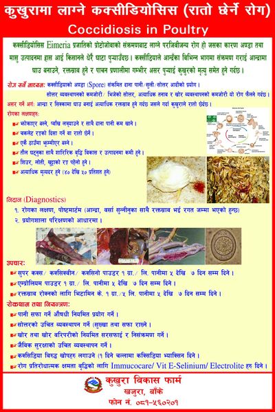
Eimeria is responsible for coccidiosis which is intracellular protozoan parasite of intestinal tract. The economically important species are E. acervulina, E. maxima, E. tenella, E. necatrix and E. brunette
Transmission
Intake of fecal contaminated food is the mode of transmission. The feces containing sporulatedEimeriaoocyst transmits the disease.
Incubation period of ceacalcoccidiosis is 5-6 days and intestinal coccidiosis is 5 days. Coccidiosis is most commonly seen in chickens between 3-6 weeks of afe although can be seen in all ages if not exposed withcoccidapreviously.young ones are more reversly affected.
⇒Cecal coccidiosis
This is most common coccidiosis caused by E. tenella. This form is characterized by profuse haemorrhage due to rupture of blood vessel. Formation of cecal core can be observed at necropsy of dead birds.
⇒Intestinal coccidiosis
E. necatrix, E. acervulina, E. mevati and E. maxima are mostly responsible for such kind of infection. Sudden death occurs at 5-7 days due to post infection. Infection leads to congestion of mucous membrane and hemorrhage. Wall of intestine becomes thick.
⇒Rectal coccidiosis
E. brunette is mostly involved in causation of this form which is characterized by lossening of wall coming out of mucous casts in droppings.
Of the seven disease producing species E. tenella, E. necatrix, E. brunette are most harmful and cause high mortality and morbidity. E. maxima and E. acervulina are moderately harmful, E. nitis, and E. preaecox are least harmful.
E. maxima, E. acervulina, and E. mitiscauses causse non haemorraghiccoccidiosis and E. tenella , E. necatrix, and E. brunette causes haemorrhagiccoccidiosis.
Clinical findings
Affected birds are apathic, dull depressed and lethargic. They pass loos dropping which may contain mucous. Some drooping may be stained with blood. Severely affected ones die and other show clinical symptoms. They are also dehydrated anemic with ruffled feather.
Lesions
Inflammation and profuse haemorrhage of intestinal tract, thickening of intestinal mucosa are found. Profuse haemorraghe in caeca in cecalcoccidiosis. haemorrhagic foci are visible on mucous membrane and caecal core are found. Ladder like haemorrahage may be seen in rectum in recalhaemorrhage.
Diagnosis
Clinical symptoms and lesions are indicative but finding of oocyst in feces is confirmatory.
Occyst of different species of coccidian are seen at following sites.
|
Coccidian |
Site |
Oocyst |
|
E. tenella |
Caeca |
Broad and ovoid, no micropyle, residuum |
|
E. necatrix |
Middle portion of SI |
Long, oval, thin wall |
|
E.acervulina |
Upper 1/3rd of SI |
Ovoid, micropyle, residuum |
|
E.maxima |
Middle and post 1/3rdof SI |
Ovoidbilayered, yellowish large |
|
E.bruneti |
Lower 1/3rdof SI |
Oval, residuum, micropyle |
|
E. imitis |
Anterior ½ of SI |
Subspherical smooth walled |
Differential diagnosis
Intestinal coccidiosis should be differentially diagnosed with necrotic enteritis.
Treatment
Sulphonamide or combination of diaverdin andsulphaquinoxaline is effective in treating coccidiosis. It is treated in specific pattern – 3 days medicated water, 2 days plain water and 2 days medicated water. Vit.A and K supplement is necessary for early recovery.
Prevention and control
Use of anti coccidial drugs on prophlctic dose controls coccidiosis. They are used on rotational and shuttle programme. Shuttle programme is the use of one anticoccidial in the starter and other in grower ration. Rotational programme is to make changes in the use of anticoccidial drugs. It is necessary to maintain good hygiene and sanitation, prevent access of infected dropping to the non-infected birds and keep older birds away from chicks, since older are carrier. Also avoid moisture and humidity in litters by frequent turning of litter to keep the litter dry to reduce the sporulation of the oocyst.
Source: https://www.farm.com.np/